
Gürkan Özkoç
Gürkan Özkoç MD FACS was born in Samsun in 1967. He graduated from Hacettepe University Faculty of Medicine in 1992. He then specialized in Orthopedics and Traumatology at Hacettepe University Faculty of Medicine. Between 1998 and 2000, he worked as an orthopedic surgeon at Private Çankaya Hospital in Ankara. In 2000, he started working at Başkent University Faculty of Medicine Adana Dr. Turgut Noyan Application and Research Center, where he served first as an Associate Professor and later as a Professor of Orthopedics and Traumatology. As of March 2020, he has been providing services to his patients under the name OrtoGrup, along with four other orthopedic professors with whom he has worked for many years.
Dr. Özkoç's professional interests include knee and hip arthroplasty, trauma surgery, orthopedic oncology, and pediatric orthopedics. He is a member of the American College of Surgeons (ACS). He is married with two children and actively enjoys cycling and long-distance running.

Ortogrup Orthopedics and Traumatology Clinic
Hurmalı Mah. Kurtuluş Cad. Central Plaza No: 39 İç Kapı No:34 Seyhan/Adana
0(322) 503 99 95 (Ortogrup Klinik)
info@gurkanozkoc.com.tr
Appointment Request
Leave
a
Comment
Leave a Comment
Our Patients' Comments

Does lactic acid accumulates in the muscles after a workout? This is an old knowledge. Post-workout muscle soreness in untrained athletes is not due to lactic acid accumulation. Muscles meet their energy needs from the anaerobic system in the absence of oxygen. Lactic acid is formed as a by-product. However, lactic acid does not accumulate in the muscles, but quickly enters the bloodstream and is converted back into glucose by the liver. Through this mechanism, called the Cori cycle, glucose is either returned to the circulation to be reused by the tissues or stored in the liver as glucogen. In this way, the energy of glucose that was not fully burned due to the lack of oxygen is put back into use. What about the pain in my muscles? (Delayed Onset Muscle Soreness (DOMS)) This condition, referred to as Delayed Muscle Soreness, is caused by the inflammation process that the body responds to micro-injuries in the muscles 48-72 hours after training.
Menisci are two C-shaped cartilage-like structures in each knee. They act as shock absorbers by cushioning the impacts on the knee. So, if they are so important, why are they removed when torn? Most meniscus tears occur in the central part, which, even if repaired, will not heal. If left as they are, these tears can grow or damage the cartilage during activities like squatting. They also cause pain. Therefore, we remove the part that cannot heal without touching the healthy part. If the rim of the meniscus remains intact, its functions are preserved, and it does not pose a significant long-term problem for the patient. On the other hand, if the tear is in the well-vascularized area near the joint capsule, we already stitch these types of tears. So, we never remove a meniscus that has a chance of healing and we repair it.
Would an MRI show abnormalities in the bones after a physically demanding sport like a marathon? A 2014 study sought to answer this question. MRIs of the hip, knee, and ankle of 16 athletes participating in national trials in the Netherlands were taken before and after the racing season and compared. The results were intriguing. Bone marrow edema was observed in 45 different regions in 14 of the 16 athletes, all of whom had no complaints. Most of these MRI findings disappeared during the season, but new ones also appeared in that time. The article notes that athletes can have various complaints during the season, but these complaints were unrelated to the MRI abnormalities. In short, if you are a runner and an MRI taken for different reasons shows bone marrow edema, it is perfectly normal. Since bone is an active tissue, bone marrow edema can be seen during the new bone formation process in response to stress. If there is no pain, this is not a reason to change your exercise plan.
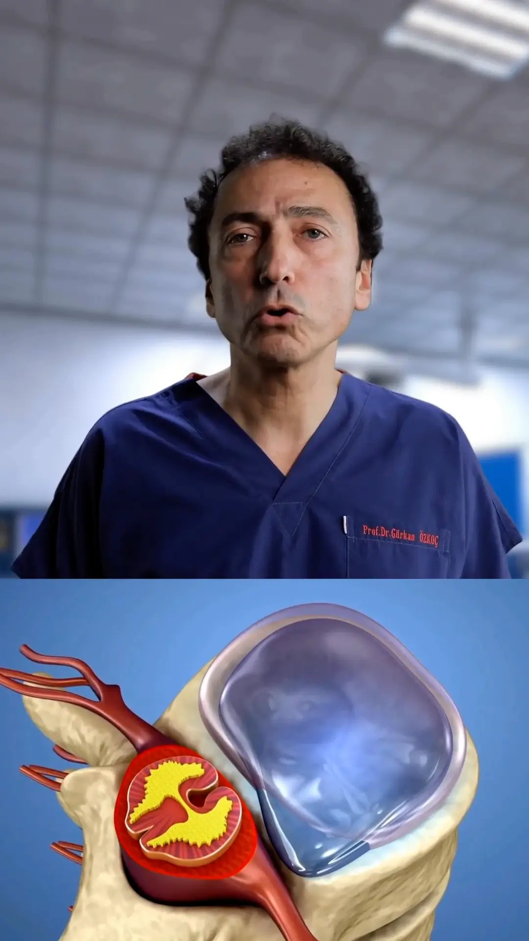
Not every mention of the meniscus in an MRI report is something to fear. Patients often perceive meniscus degeneration noted in an MRI taken for other reasons as a tear, and they may even think surgery is necessary. Menisci are structures that act as cushions in load transfer between the knee bones. While their internal structure is completely healthy in the twenties, over the years, and especially with increasing weight, internal structural deteriorations can occur, as seen here. Sometimes, even without excess weight, degeneration can also occur after repetitive microtraumas and long-term sports like football. Stage I and II degenerations are not tears and do not require surgery. However, when Stage III is mentioned, it indicates a real tear. Even then, not every meniscus tear requires surgery, so it is best to follow your doctor's recommendations.
Leg pains in children are often dismissed by families and doctors as growing pains. But which ones are truly growing pains? Contrary to popular belief, growing pains are seen between the ages of four and six, not during the rapid growth period of 10 to 14 years. So, which pains in children are important?
Growing pains typically occur outside the joints and in the legs, are symmetrical, usually happen in the evening or throughout the night after a busy day, and disappear by morning. They do not involve symptoms like increased warmth, redness, or swelling and are generally temporary, with laboratory and radiological studies mostly negative.
On the other hand, pains that are unilateral and persistent, occur in the morning and continue throughout the day, are accompanied by redness, warmth, and swelling, are progressively worsening, and show abnormal findings in lab tests and X-rays should definitely be taken seriously.
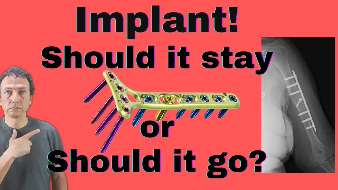
Fracture healing is a natural process. We know that animals' bones can heal on their own in the wild. So, why do we perform surgery when a fracture occurs?
-To ensure the original length, rotation, and alignment of long bones, and to perfectly restore the joint line in intra-articular fractures since even a few millimeters of step-off in the cartilage can lead to osteoarthritis in later years.
-To provide the immobility needed at the fracture ends for healing using plates, screws, and nails
- To stabilize fractures that would take a very long time to heal with a cast, preserving joint movement and muscle strength.
-To eliminate pain associated with movement by surgically stabilizing the fracture ends.
-To speed up fracture healing by allowing the implanted hardware to bear the load.
-To repair additional injuries such as vascular or nerve damage.
Sometimes, questions like "I had surgery for a fracture in a certain area; when will it heal?" are asked on social media. The healing time of a fracture depends on so many factors. It varies greatly depending on which bone is fractured, whether it is at the end or the middle of the bone, the nature of the trauma and the magnitude of the energy burst in the area, the condition of the soft tissue damage, which is the most important factor in bone nutrition, the patient’s comorbidities and medications, nutritional status, age, smoking habits, the success of the surgery, and whether early rehabilitation is performed.
For patients I treat, I provide an expected healing time based on our treatment. This period can also vary greatly. On the other hand, making a comment without knowing all these variables is no different from reading coffee grounds.
MRI, or Magnetic Resonance Imaging, uses magnetic fields and radio waves to provide detailed information about tissues. Contrary to what some patients believe, it does not involve radiation and is extremely safe. However, three factors should be considered:
1. The powerful magnetic field of the MRI machine can move or heat metal objects in the body. Items like pacemakers, certain vascular stents, inner ear implants, and some very old prostheses may contain non-MRI-compatible metals, so these should be reviewed before the scan.
2. Since the effects of MRI on pregnancy are not fully understood, MRI is generally not recommended during the first trimester of pregnancy. However, it can be performed when considering the risks and benefits.
3. For patients with claustrophobia, MRI can be administered with sedative medications.
